When I teach service members without a medical background, I explain the nasopharyngeal airway through anatomy: the tube passes through the nose into the pharynx and may provide a route for air behind the tongue base. A diagram makes this understandable. But it shows an idealised position. In an actual casualty, the tip is not visible, and an external measurement cannot confirm that the device has reached the position we expect.
This raises a teaching question: do we spend enough time explaining how to assess whether an NPA helps and recognise when it is insufficient? Insertion is only part of the intervention. The next part begins by assessing what has changed in the casualty’s breathing.
What an airway adjunct can change
An NPA is an adjunct for maintaining upper-airway patency. Reduced muscle tone can allow soft tissues to narrow the air passage. The tube may help bypass some of that narrowing, but its effect depends on its relationship to those tissues. Tseng et al., 2019.
Four functions should be distinguished. Patency means that air can move through the airway. Ventilation moves air into and out of the lungs, including the removal of CO₂. Oxygenation supplies oxygen to the blood. Protection against aspiration prevents blood, vomit or other material from entering the lower airway. These processes are related, but they are not interchangeable.
Traumatic obstruction or increasing swelling may exceed what an NPA can address and require timely assistance from an appropriately trained clinician. An NPA does not generate breaths, repair injured lungs or seal the entrance to the trachea. Its presence therefore does not establish adequate ventilation or protection against aspiration. Low oxygen saturation does not prove an upper-airway problem either: breathing and other possible causes of deterioration must be assessed. TCCC, 1 May 2026.
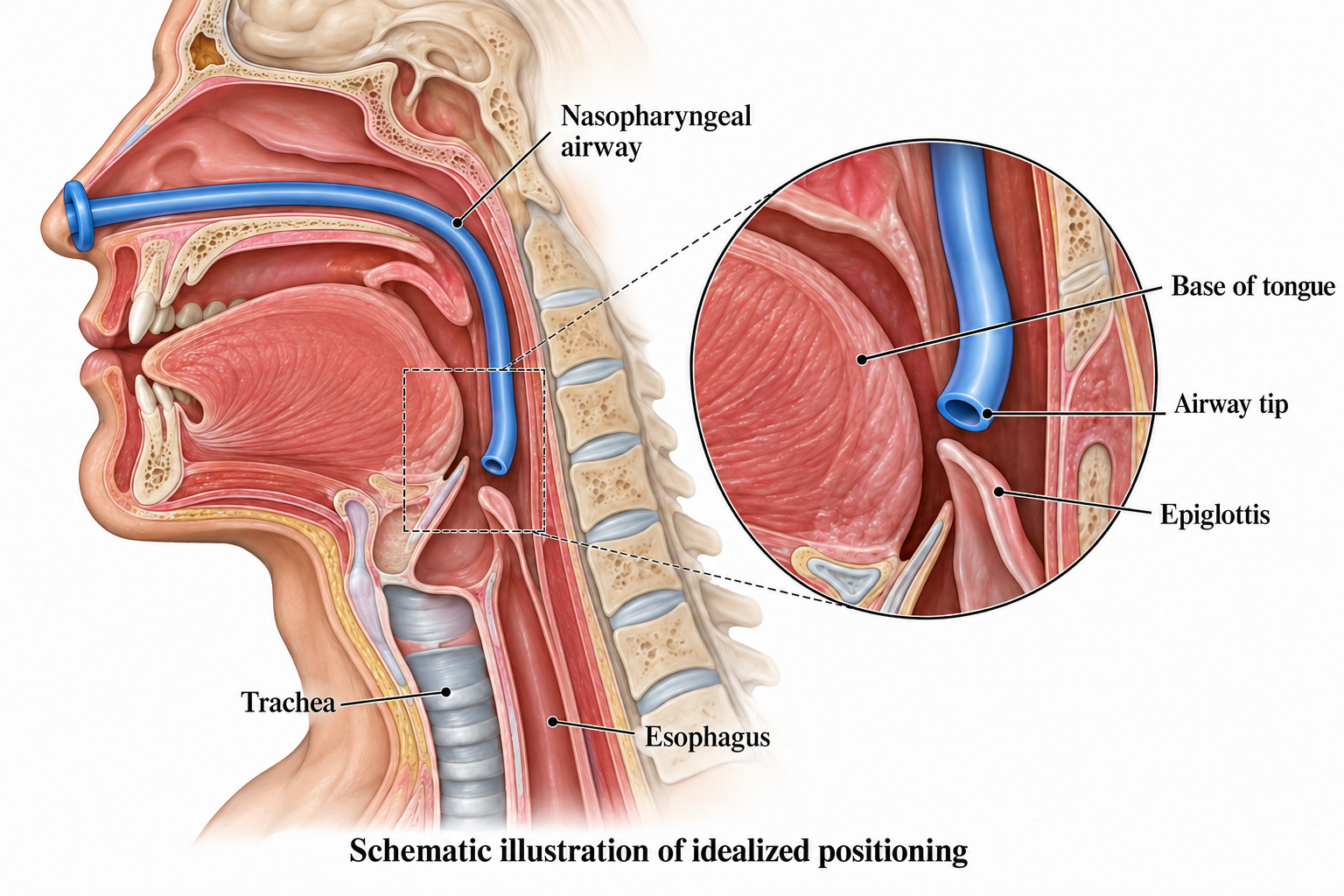
This illustrative diagram explains the expected position; it does not confirm placement in an individual casualty.
Sizing informs selection, not verification
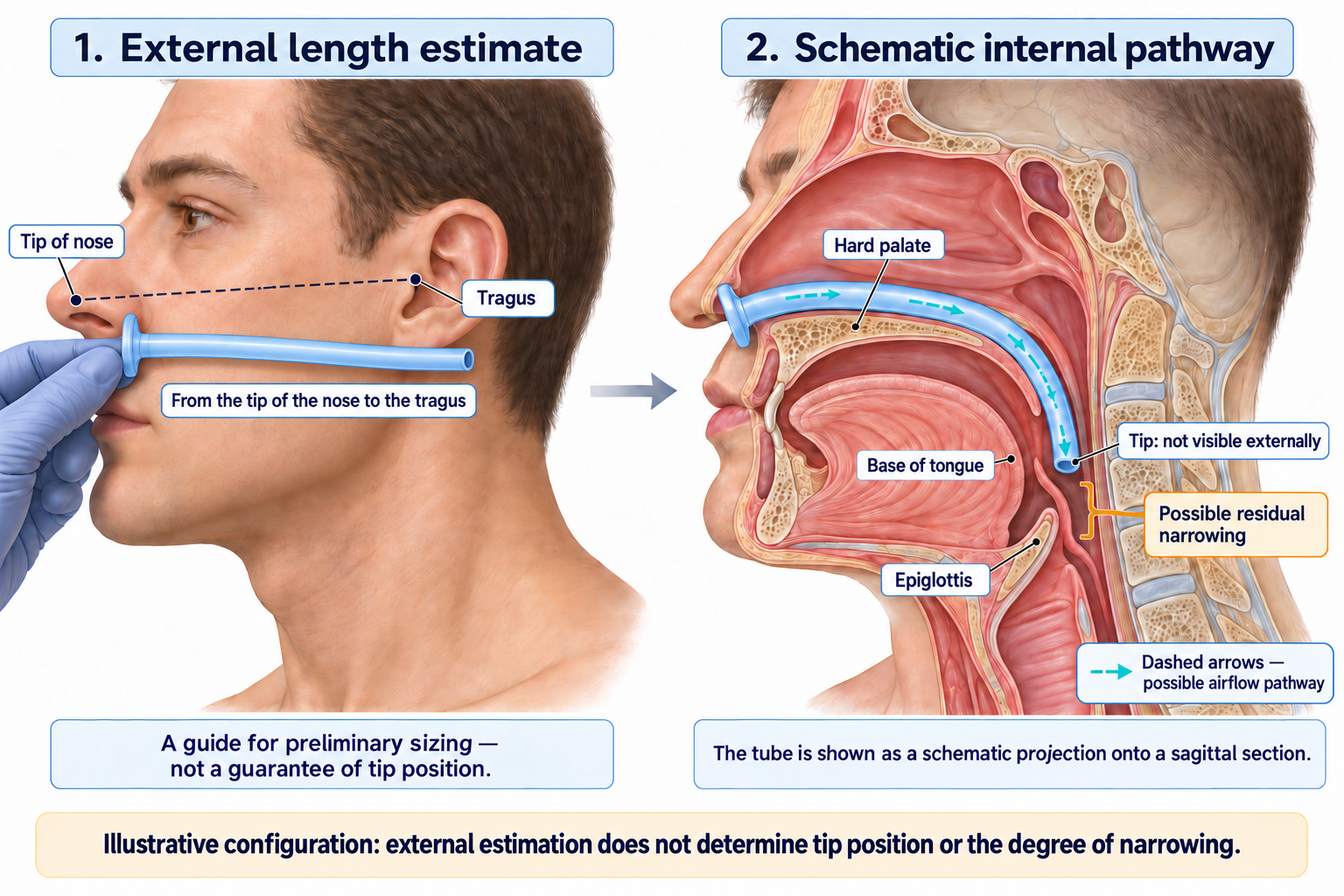
Length matters: the tip needs to reach the region where the tube can assist airflow. A device that is too short may fail to bypass narrowing; excessive length may place the tip in an unintended location. Diameter also matters for passage through the nose, but it cannot substitute for considering length.
External landmarks help with initial selection. Their limitation is that they estimate an internal distance indirectly. Facial shape, pharyngeal anatomy and traumatic changes differ between people. Matching a landmark provides neither a view of the tip nor evidence of unobstructed airflow.
This does not make measurement pointless. Tseng and colleagues found relationships between external measurements and internal distance, but restricted the applicability of their findings to the population studied and excluded patients with anatomical abnormalities of the face and neck. Full study.
Head position remains part of the assessment. An NPA does not make casualty positioning irrelevant. Breathing needs reassessment after repositioning or transfer. Positioning and manoeuvres should account for the injury and applicable protocol, rather than mechanically reproduce a diagram.
What the evidence actually shows
In 1993, Stoneham assessed NPA position and performance in 120 anaesthetised adults using fibreoptic laryngoscopy. Findings included malposition, tube compression and soft-tissue obstruction. Three external measurements did not significantly correlate with the nares-to-epiglottis distance in that sample. These findings are reported in the author’s abstract the full text was unavailable when this article was prepared.
This demonstrates that an inserted NPA may not relieve obstruction. It does not establish a battlefield failure rate. Anaesthetised patients and combat casualties differ in tissue conditions, injuries and circumstances of care.
Tseng’s anatomical measurements inform a discussion of length; they do not establish a survival benefit. Manikin simulation can support practice of technique and decisions, but cannot reproduce every property of a living pharynx. Successful performance on a model is not clinical evidence of effectiveness.
The TCCC change paper highlights limited prehospital NPA evidence. Small numbers of recorded battlefield uses cannot establish either benefit or a reliable failure rate. Insufficient evidence is not proof of ineffectiveness. Shaw et al., 2024.
What changed in TCCC
Change 24-1 in 2024 removed routine NPA use for an unconscious casualty without traumatic obstruction from the airway section. The emphasis shifted to positioning, keeping the chin away from the chest, suction when available, and an appropriate response to traumatic obstruction. The NPA remained a possible adjunct to bag-mask ventilation. Shaw et al., 2024.
In the 1 May 2026 edition, section 5e advises considering a properly sized NPA and ventilation with a 1000-mL resuscitator bag-valve-mask when ventilation is impaired and hypoxia cannot be corrected, with decreasing SpO₂ below 90%, or below 92% with moderate/severe TBI. Section 5f calls for continuous EtCO₂ and SpO₂ monitoring to help assess patency. Bag capacity is not the volume to deliver with every breath. TCCC 2026, section 5.
This is a change in the device’s role, not a complete prohibition. The guideline wording does not establish removal from every kit or training level. Nor do these thresholds mean that obvious ineffective breathing should go untreated while waiting for a monitor to display a particular number.
A bag-mask does not remove the need to assess
A bag generates pressurised airflow; it does not guarantee delivery to the lungs. If the chest does not rise appropriately, reassess patency, positioning, mask seal and other causes of ineffective ventilation within the provider’s training. Squeezing harder is not a solution to “push through” obstruction: excessive ventilation can contribute to gastric inflation and regurgitation. AHA, Adult Basic Life Support, 2025.
Visible chest movement is useful, but not conclusive. Consider a hypothetical example: airway noise decreases, yet breaths remain shallow and consciousness deteriorates. That is insufficient evidence that “the NPA is working.” In another example, the chest moves but saturation falls. The assessment must extend beyond the tube’s position.
Teaching what happens after insertion
A non-medical responder can compare the casualty before and after the intervention: is airflow detectable, is breathing less laboured, what are the depth and rhythm of breaths, how do the chest and abdomen move, and is consciousness changing? Snoring, gurgling, paradoxical movement or weak airflow despite respiratory effort should prompt immediate reassessment, rather than a completed “airway inserted” checkbox. These signs need demonstration and explanation. Resuscitation Council UK, ABCDE.
Silence alone is not success: complete obstruction may leave no airflow to hear. A medic adds monitoring, assessment of signal quality and trends. SpO₂ does not measure ventilation and may be misleading in shock or hypothermia. EtCO₂ helps assess exhaled carbon dioxide, but does not reveal the exact location of an NPA tip. Monitor findings must be interpreted alongside the casualty’s condition. TCCC 2026, RCUK.
For a teaching scenario, I propose five questions:
- What problem did we expect to change?
- What signs were present beforehand?
- What actually changed afterwards?
- What indicates that this intervention is insufficient?
- What next action is within our training, and whose assistance is needed?
Reassessment belongs immediately after the intervention, after movement and whenever deterioration occurs. “Every five minutes” is not a universal NPA rule or permission to leave an unstable casualty unobserved. Failure to improve requires timely medical assistance, continued measures within training and arrangements for further care. It does not authorise a non-medical responder to perform more advanced procedures.
For instructors, insertion, assessment of effect and an appropriate response to failure should all be included in the scenario. This article explains that reasoning; it does not replace practical training. The outcome to assess is whether breathing has become more effective and remains so, alongside the quality of the insertion technique.
References
- Shaw T, Grisham J, Kotora J, et al. Airway Management in Tactical Combat Casualty Care: TCCC Change 24-1. Journal of Special Operations Medicine. 2024;24(4):45–56. Full PDF.
- Committee on Tactical Combat Casualty Care. Tactical Combat Casualty Care Guidelines. 01 May 2026. NAEMT PDF.
- Stoneham MD. The nasopharyngeal airway. Assessment of position by fibreoptic laryngoscopy. Anaesthesia. 1993;48(7):575–580. doi:10.1111/j.1365-2044.1993.tb07119.x. PubMed — abstract checked.
- Tseng WC, Lin WL, Cherng CH. Estimation of nares-to-epiglottis distance for selecting an appropriate nasopharyngeal airway. Medicine (Baltimore). 2019;98(10):e14832. doi:10.1097/MD.0000000000014832. Full text.
- American Heart Association. Part 7: Adult Basic Life Support. 2025 Guidelines for CPR and ECC. Official text.
- Resuscitation Council UK. The ABCDE Approach. Updated July 2024. Official text.