Introduction
The mechanism of action of kaolin is often explained in a single sentence:
“Kaolin activates Factor XII and initiates the intrinsic coagulation pathway.”
Formally, this is correct. But this explanation is not enough to understand why hemostatic gauze works in a real wound — and why even the correct hemostatic agent sometimes fails to stop bleeding.
Control of massive hemorrhage depends not only on the hemostatic agent itself, but also on where, how, and under what conditions it is applied.
Kaolin does not “seal” a damaged vessel on its own. It does not glue tissues together, create an airtight plug, or affect the bleeding source if the hemostatic gauze is not physically in contact with it. Its role is different: to create local conditions in which the casualty’s own hemostatic system can form a stable clot more quickly.
That is why, to understand Combat Gauze, it is important to separate two processes:
mechanical bleeding control — wound packing and compression;
biochemical enhancement of hemostasis — the effect of kaolin.
Without the first, the second may be almost useless.
What is kaolin?
Kaolin is a naturally occurring mineral material based on kaolinite, an aluminosilicate with pronounced surface properties.
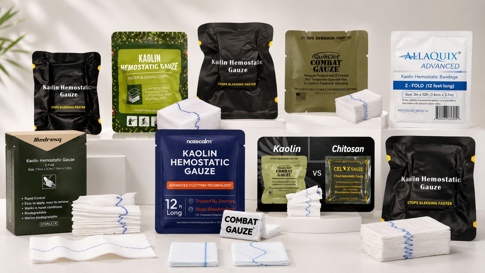
In hemostatic products such as QuikClot Combat Gauze, kaolin particles are applied to the gauze. Contact between blood and this mineral surface initiates what is known as contact activation of the coagulation system. The electrical charge of the kaolin surface is particularly important.
Negatively charged surfaces can promote activation of Factor XII (FXII, Hageman factor). This phenomenon has been known for a long time and is used, among other things, in laboratory coagulation tests. Experimental studies demonstrate contact activation of FXII on kaolin-containing surfaces (2, 3). These are mechanistic laboratory findings, not evidence of the clinical superiority of a particular dressing.
In other words, kaolin does not contain ready-made thrombin, fibrin, or other clotting factors.
It does not add a new hemostatic system to the body.
Instead, it creates an active surface on which the casualty’s own blood components can react more rapidly in ways that promote clot formation.
This is what fundamentally distinguishes kaolin, for example, from materials whose mechanisms rely largely on direct interaction with blood cells or on the mechanical formation of a barrier.
How does the mechanism work?
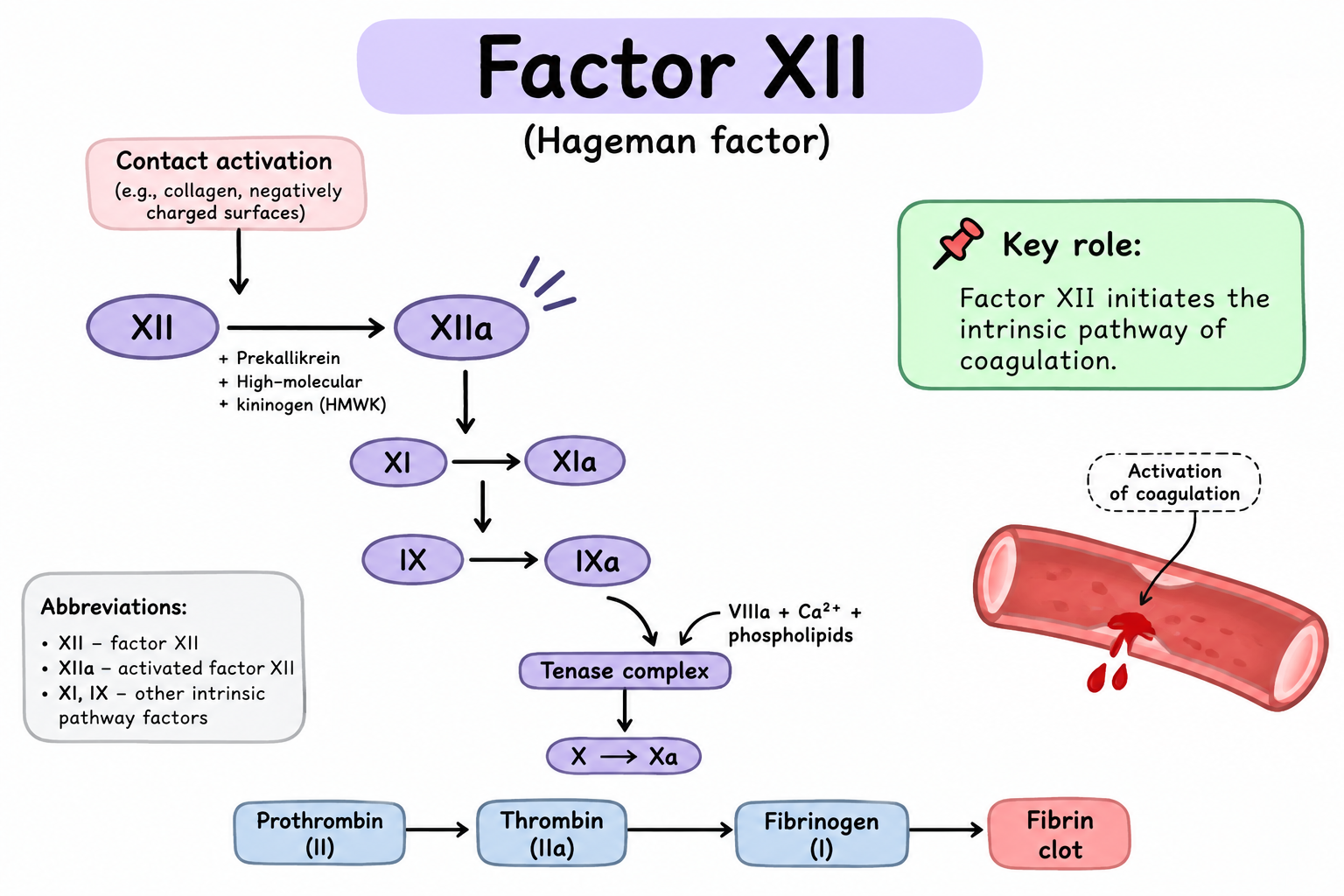
In the classical coagulation cascade, the mechanism can be described approximately as follows:
kaolin → activation of XII → XI → IX → X → thrombin generation → conversion of fibrinogen to fibrin.
More precisely, activated Factor XII (FXIIa) promotes activation of Factor XI. Then, with the participation of Factors IX and VIII, activation of Factor X is amplified. Factor Xa, together with Factor Va, converts prothrombin into thrombin, while thrombin subsequently converts fibrinogen into fibrin and participates in numerous positive feedback loops within the coagulation system. The result is faster formation of the fibrin component of the clot.
This is the origin of the common explanation:
“Kaolin activates the intrinsic coagulation pathway.”
But this is where things become more interesting.
Why is Factor XII alone not enough to explain it?
There are people with severe inherited Factor XII deficiency. Their laboratory tests may show a markedly prolonged aPTT (activated partial thromboplastin time). Logically, one might expect such a person to experience severe bleeding. But this usually does not happen.
Even profound congenital FXII deficiency, unlike deficiencies of Factors VIII or IX, does not cause the typical clinical bleeding phenotype (5). This is one of the classic examples of how the laboratory model of the “intrinsic” and “extrinsic” pathways differs from hemostasis as it actually occurs in vivo.
The traditional coagulation cascade remains very useful for teaching and for understanding laboratory tests. However, true hemostasis in injured tissue is considerably more complex. The modern cell-based model of hemostasis views coagulation not as two independent tubes — “intrinsic” and “extrinsic” — that simply converge on Factor X, but as an interaction of reactions occurring on the surfaces of different cells.
In real trauma, the following interact simultaneously:
- tissue factor;
- activated platelets;
- coagulation factors;
- thrombin and its positive feedback loops;
- cellular phospholipid surfaces;
- fibrinogen;
- endothelium;
- the fibrinolytic system;
- local blood flow;
- temperature;
- pH;
- concentration and availability of coagulation factors.
The cell-based model therefore explains hemostasis in vivo better than a simple diagram of two independent coagulation cascades (4).
For this reason, the role of kaolin is better described not as: “Kaolin initiates blood clotting.”
But rather as:
“Kaolin creates an artificial procoagulant surface at the site of bleeding and enhances local clot formation.”
And that distinction is fundamental. Kaolin amplifies the casualty’s hemostatic system, but it does not replace it. It is not a tissue adhesive. It does not create an independent physical seal over the damaged vessel. It does not compress the vessel. And it cannot act on the source of bleeding merely because the hemostatic gauze is somewhere inside the wound. Contact is required.
Practical significance
This is where biochemistry becomes practical tactical medicine. The real mechanism of bleeding control does not look like this:
kaolin → bleeding stopped.
It looks more like this:
contact with the source of bleeding
→ tight wound packing
→ elimination of dead space
→ continuous compression
→ sufficient time
→ clot formation and stabilization.
And it is only within this process that kaolin does its job.
Wound packing
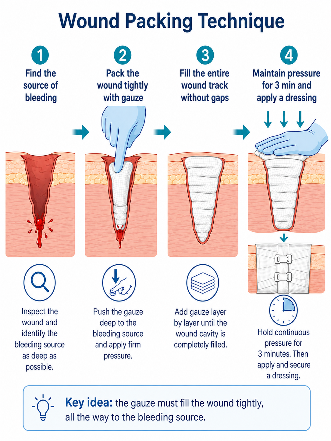
The main purpose of wound packing is to deliver the hemostatic material directly to the source of bleeding. This means more than simply filling the entrance of the wound tract with gauze. You need to reach the deepest point of the wound, identify the area of most intense bleeding, and begin packing from that point.
By tightly filling the cavity, we simultaneously:
- create direct contact between the material and blood;
- reduce the space into which blood can continue to accumulate;
- create mechanical resistance to blood flow;
- create conditions for local clot formation.
If the hemostatic gauze lies only near the entrance of the wound tract while the damaged vessel is much deeper, the presence of kaolin alone does not solve the problem.
Compression
The next component is direct pressure.
Compression temporarily reduces blood flow through the injured area and allows the clot to form and remain in place. Without adequate compression, high blood flow can mechanically interfere with formation of a stable clot or disrupt it before it becomes sufficiently strong.
That is why an active hemostatic agent does not eliminate the need for direct pressure. The May 1, 2026 TCCC Guidelines recommend Combat Gauze as the CoTCCC hemostatic dressing of choice for external compressible hemorrhage that is not anatomically amenable to an extremity tourniquet, as well as an adjunct during tourniquet conversion. Combat Gauze should be applied with at least three minutes of direct pressure (1).
When should you think about kaolin?
Primarily in cases of massive external compressible hemorrhage that is not anatomically suitable for conventional tourniquet application.
A typical example is a deep wound in a junctional area or another location that can be effectively packed. Hemostatic gauze may also be used as part of controlled tourniquet conversion when the appropriate TCCC criteria are met and bleeding can be safely controlled by other methods. At the same time, the presence of Combat Gauze should never delay tourniquet application when a tourniquet is indicated.
If there is massive extremity hemorrhage that can be rapidly and effectively controlled with a recommended tourniquet, TCCC calls for tourniquet application.
When should you not expect kaolin to solve the problem?
Hemostatic gauze does not compensate for choosing the wrong bleeding-control method.
It will not rescue the situation if:
- a tourniquet is indicated but its application is delayed;
- the source of bleeding cannot physically be reached by wound packing;
- the gauze fills only the superficial part of the wound while the bleeding source remains deep;
- the wound is not packed tightly enough;
- direct pressure is weak or repeatedly interrupted;
- the clot is not given enough time to form;
- bleeding control is not reassessed after casualty movement or during evacuation.
In such a situation, “hemostatic gauze” can easily become nothing more than very expensive gauze. And the problem will not be a lack of biochemical activity of kaolin, but rather that the conditions required for that activity to have practical significance were never created.
What about traumatic coagulopathy?
Here too, it is important to avoid an overly simplistic explanation. Because kaolin uses and enhances the casualty’s own coagulation system, it is entirely logical that the condition of that system matters.
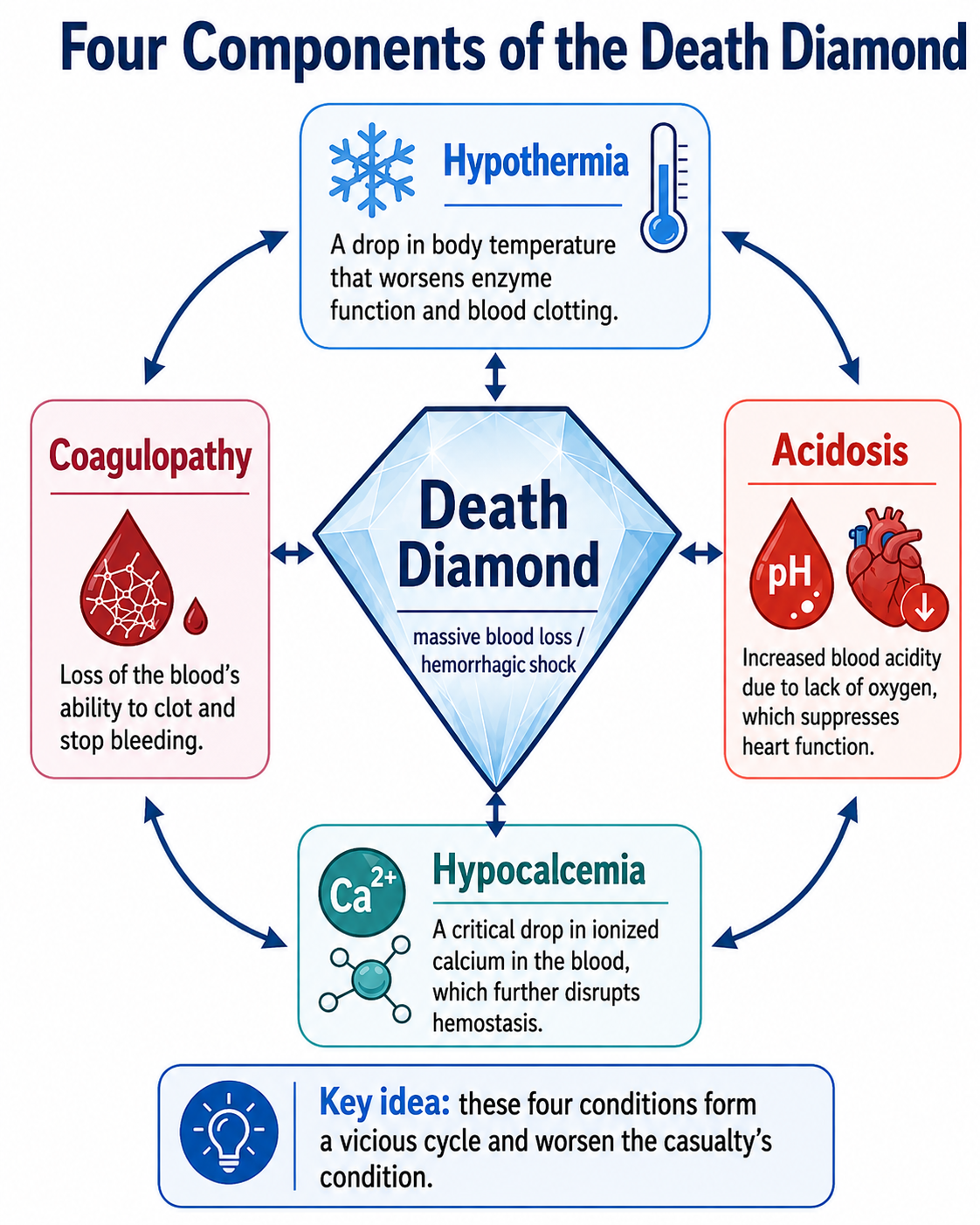
Severe blood loss may be accompanied by:
- hypothermia;
- acidosis;
- hemodilution;
- reduced fibrinogen concentration;
- coagulation factor deficiency;
- impaired platelet function;
- pathological changes in fibrinolysis.
All of these can make formation of a stable clot more difficult. But this does not mean that “kaolin does not work in coagulopathy.” That would be an equally excessive simplification.
Experimental animal studies have demonstrated the effectiveness of kaolin-containing hemostatic materials even under conditions of significant hemodilution, hypothermia, acidosis, and laboratory-confirmed coagulopathy. For example, Combat Gauze has been studied in hypothermic and coagulopathic models of severe liver injury, in a bleeding model involving hemodilution and hypothermia, and in a modern junctional hemorrhage model with acidosis to approximately pH 7.2 and coagulopathy (6, 7, 8).
At the same time, these studies are predominantly experimental animal studies, so they should not be interpreted as evidence that severe traumatic coagulopathy ceases to matter in a real casualty.
The correct conclusion is different:
physiological impairment of hemostasis can reduce the effectiveness of any bleeding-control strategy, but this does not mean an automatic loss of kaolin’s effect.
That is why bleeding control cannot be separated from hypothermia prevention, appropriate resuscitation, shock management, and repeated reassessment of the casualty.
Key practical takeaway
The mechanism of action of a hemostatic agent should help us make decisions. But it cannot replace correct technique. Kaolin does not replace wound packing.
It enhances the result of properly performed wound packing by accelerating clot formation precisely where contact with the bleeding source and adequate compression have already created the necessary conditions.
Perhaps this is the best sentence for teaching Combat Gauze: Kaolin does not pack the wound for you. It makes correctly performed wound packing more effective.
References
Joint Trauma System / Committee on Tactical Combat Casualty Care. Tactical Combat Casualty Care Guidelines — official Deployed Medicine page. Edition used: May 1, 2026, Tactical Field Care, section 4: Massive Hemorrhage. The date was checked against the original PDF title page. Massive Hemorrhage section: Combat Gauze is designated as the CoTCCC hemostatic dressing of choice for compressible external hemorrhage not amenable to an extremity tourniquet and as an adjunct to tourniquet removal; Combat Gauze is applied with at least 3 minutes of direct pressure.
Zhu S, Diamond SL. Contact activation of blood coagulation on a defined kaolin/collagen surface in a microfluidic assay — PubMed. Thrombosis Research. 2014;134(6):1335–1343. The study demonstrates activation of the contact system and FXII on a kaolin-containing surface, followed by platelet deposition and fibrin formation.
Griffin JH. Role of surface in surface-dependent activation of Hageman factor (blood coagulation factor XII) — PubMed. Proceedings of the National Academy of Sciences. 1978;75(4):1998–2002. A classic study on the role of negatively charged surfaces, including kaolin, in contact activation of FXII.
Hoffman M, Monroe DM. A Cell-based Model of Hemostasis — PubMed. Thrombosis and Haemostasis. 2001;85(6):958–965. One of the key papers that shaped the modern cell-based understanding of hemostasis and demonstrated the limitations of the classical “intrinsic/extrinsic pathway” model for describing coagulation in vivo.
Fernandes HD, et al. Factor XII Deficiency Mimicking Bleeding Diathesis: A Unique Presentation and Diagnostic Pitfall — PubMed. Cureus. 2018;10(6):e2817. A case report of FXII deficiency with markedly prolonged aPTT but no characteristic clinical bleeding tendency.
Johnson D, Bates S, Nukalo S, et al. The effects of QuikClot Combat Gauze on hemorrhage control in the presence of hemodilution and hypothermia — PubMed. Annals of Medicine and Surgery. 2014;3(2):21–25. An experimental study of Combat Gauze effectiveness under conditions of hemodilution and hypothermia.
Sena MJ, Douglas G, Gerlach T, et al. A pilot study of the use of kaolin-impregnated gauze (Combat Gauze) for packing high-grade hepatic injuries in a hypothermic coagulopathic swine model — PubMed. Journal of Surgical Research. 2013;183(2):704–709. A study of kaolin-impregnated gauze in a severe liver injury model under conditions of hypothermia and coagulopathy.
Gerling KA, Kersey AJ, Lauria AL, et al. Evaluation of novel hemostatic agents in a coagulopathic swine model of junctional hemorrhage — PubMed. Journal of Trauma and Acute Care Surgery. 2023;95(2S):S144–S151. In a model of severe coagulopathy and acidosis, Combat Gauze was used as the military standard control for comparison with contemporary hemostatic agents.
Revision date: